Phase Information of the MR Signal
Magnetic resonance imaging (MRI) is a tremendously successful tool for biomedical research and diagnostic imaging. MRI research resides at the interfaces of life sciences and natural sciences and their respective subdisciplines.
Most of the information gathered with MRI is due to subtle variations in signal magnitude, which can be translated into images with meaningful information about the nuclei’s biophysical environment. The MRI signal, however, has another, promising, yet underexploited property: phase. The phase is best explained by using the analogy of a lighthouse: The MRI signal’s magnitude corresponds to the brightness of the lighthouse beam and the MRI signal’s phase corresponds to the beam’s direction. Just as the lighthouse beam always has a certain direction, the MR signal always has a certain phase. One challenge with phase is that we do not yet fully understand the underlying biophysical processes leading to phase information. The conventional contrasts have been investigated for decades and are, although still not fully understood, used extensively in MRI. Phase may allow us to glean further information on tissue composition and structure.
MR Data Processing
We have developed a region growing algorithm that uses a combination of data quality criteria extracted from the complete complex information. In MRI this is important is we want to preserve all spatial frequencies. Temperature mapping or maps of the magnetic field are examples for such applications. The classic approach in phase imaging, homodyne, filtering does not preserve all spatial frequencies. Usually homodyne filtering is good enough, but in some situations phase unwrapping is superior. One example is venography in areas with strong field inhomogeneities. Another example is mapping of the R2* decay, for example with multi echo SWI, where the correction for unwanted signal decay due to background field inhomogeneities requires field maps.
Applications
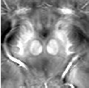
We use the phase’s sensitivity to iron for the imaging of iron rich brain strcutures. This allows us the visualization of areas that are difficult to image with conventional MRI. One example is the subthalamic nucleus subthalamic nucleus, which is a target of deep brain stimulation in Parkinson Disease. It was shown that iron is elevated in deep gray matter structures of the brains of people with multiple sclerosis or Parkinson disease. We use our multi echo SWI technique to map these structures.